


Discoveries Magazine Logo
From Wearable Sensors to Printed Placentas
Maternal and fetal health researchers team up with engineers to improve pregnancy outcomes
Story by:
- Susanne Clara Bard
–
scbard@ucsd.edu


Explore the 2026 IssueDiscoveries Magazine
Published Date
August 19, 2026
Story by:
- Susanne Clara Bard
–
scbard@ucsd.edu
Explore the 2026 IssueDiscoveries Magazine
Article Content
This story is from the 2026 issue of Discoveries, a UC San Diego Health Sciences magazine
As a first-timeparent-to-be, 30-year-old Sarah Cleveland wanted an unmedicated vaginal delivery but felt nervous about it. “I don’t know anyone who has given birth without medication,” she said. “I felt like I needed to do something to prepare.”
Her anxiety was justified. Pelvic floor disorders stemming from lacerations and nerve damage during delivery are a little-known but painful reality for some birthing parents, often resulting in incontinence, chronic pain, pelvic organ prolapse and sexual dysfunction
“Even though they’re not life‑threatening, these disorders are debilitating,” saidLindsey Burnett, MD, PHD, assistant professor in the Department of Obstetrics, Gynecology and Reproductive Sciences (OBGYN & RS) in the Division of Urogynecology and Reconstructive Pelvic Surgery at UC San Diego School of Medicine and director of the Pelvic Health After Birth Program at UC San Diego Health. “Pelvic muscles get stretched to over 300% of their resting length during vaginal delivery; any other muscle in your body would be irreversibly damaged.”
According to Burnett, very little is known about the biology of pelvic floor disorders and how to prevent them, so she teamed up with Ken Loh, PHD, TaylorMade Golf Chancellor’s Endowed Faculty Fellowship in Structural Engineering at UC San Diego Jacobs School of Engineering, to investigate whether progressive weight‑bearing exercise can protect the pelvic floor. The researchers received a 2025 Galvanizing Engineering in Medicine award from the Institute of Engineering in Medicine for their Biomechanical Monitoring for Birth Injury Prevention pilot study.
When Cleveland heard about it, she signed up right away
Pelvic floor disorders, like many pregnancy‑related conditions, have historically received little research attention
“Women’s health care has traditionally been underfunded, but UC San Diego is leading innovation to ensure perinatal health gets the focus it deserves,” said OBGYN & RS department chair Cynthia Gyamfi-Bannerman, MD, MS
Burnett and Loh’s study is one of several collaborations between UC San Diego OBGYN & RS and engineering researchers using the power of technology to help bridge the knowledge gap
Stretching the limits
Knowing how detrimental pelvic floor disorders can be, Burnett worked out with weights throughout her own two pregnancies and gave birth with no tearing. She wondered whether weight-bearing exercise can have the same effect on other pregnant women
“Data suggests exercise during pregnancy has many benefits,” Burnett said. “The question is what kind of exercise is feasible, and how much do you need to make a difference?”
Beginning in the second trimester, Cleveland and her fellow participants work out at a local gym three times a week for 6 weeks under the guidance of professional prenatal fitness trainers
“There are a lot of stretches and weighted exercises aimed at strengthening our core and our pelvic floor,” she said
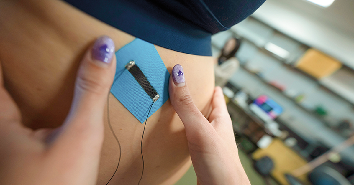
Loh’s lab develops “smart” materials to accurately measure human performance in real time. For the study, his team created wearable sensors by printing nanomaterials directly onto commercial kinesiology tape, which adheres to the inner thigh, glutes and back to track muscle engagement during the exercise sessions
The sensors, called Motion Tape, detect minute skin deformations that correlate with muscle contraction and exertion and transmit the data wirelessly to a phone app or laptop
“Motion Tape is extremely versatile and can be placed anywhere on the body, such as the chest region to measure breathing. We also use a camera‑based motion‑capture system to document the 3D movement of participants — data that can be used to optimize exercise regimens,” said Loh
The pilot study is following participants for 6 weeks postpartum, tracking cesarean versus vaginal delivery rates as well as the impact of the exercises on perineal tearing and pelvic floor strength
Burnett and Loh hope to expand the study to more participants, with the ultimate goal of providing clinicians with data‑driven guidelines for effective prenatal exercise programs. If the sensor‑guided workouts prove effective, they can potentially reduce birth‑related injuries and empower patients such as Cleveland to approach childbirth with confidence
Injecting hope
While prevention is the ultimate goal, better treatments for pelvic floor injuries are also urgently needed
“There are huge gaps in postpartum health research,” said OBGYN & RS ProfessorMarianna Alperin, MD. “We don’t know how pelvic skeletal muscles recover from injury because almost all of the research is on male animals or leg muscles.”
Alperin co‑directs the UC San Diego Center for Women’s Health Innovations through Scientific Discoveries, Engineering, and Medicine, a research hub that connects engineers, basic scientists and clinicians
“The institution is extremely conducive to collaborative efforts between engineers who make technology and clinicians who know what women need,” she said. “That’s what makes UC San Diego unique.”
Alperin has collaborated with Pierre Galletti Endowed Chair for Bioengineering InnovationKaren Christman,PHD, to develop and test an injectable hydrogel made from muscle-tissue derived extracellular matrix (ECM) to repair the pelvic floor. The hydrogel is one of many biomaterials developed by Christman’s research group. ECM is the 3D framework of proteins and carbohydrates that surrounds tissues and organs.
Remote monitoring offers a way to protect families during those critical weeks at home.
“The hydrogel acts as a scaffold that recruits the body’s own cells and encourages new muscle formation,” said Christman, who co-directs the Sanford Advanced Therapy Center at the Sanford Stem Cell Institute
In an animal model of simulated birth injury, injecting the hydrogel directly into pelvic floor muscles promoted muscle regeneration and reduced scar tissue. The team plans to seek approval from the FDA to conduct rigorous clinical trials of the hydrogel in postpartum women who show signs of pelvic floor muscle dysfunction — potentially offering a minimally invasive alternative to surgery
Predicting preeclampsia
While pelvic floor injuries can be debilitating, preeclampsia — high blood pressure during or after pregnancy — is a leading cause of maternal and fetal morbidity and mortality, according to Assistant Professor of OBGYN & RS Marni Jacobs, PHD ‘13
“Preeclampsia is a very severe complication of pregnancy,” she said
The condition currently affects up to one in 12 pregnancies in the U.S. but is on the rise. It can lead to preterm birth, stillbirth, maternal organ damage and long‑term cardiovascular disease. Complications and death due to preeclampsia can be prevented with better monitoring and treatment, but it remains difficult to diagnose and even harder to predict, Jacobs said
Researchers in the School of Medicine’s Division of Maternal and Fetal Health aim to change that through a variety of research initiatives. They’re increasingly embracing emerging technologies, from wearable biometric monitors to 3D bioprinted models of the human placenta, to identify early signs of the disorder
An early warning system
To improve the detection of postpartum preeclampsia and other cardiovascular complications, Assistant Professor of OBGYN & RSUkachi Emeruwa, MD, MPH, launched the WATCH Postpartum Hypertension study, a clinical trial that sends new parents home with a smartwatch equipped with an inflatable blood pressure cuff
With a single press of a button, the cuff inflates, captures a blood pressure reading and automatically transmits the data to the patient’s electronic medical record. Participants take eight readings a day for 6 weeks to detect the large fluctuations characteristic of postpartum cardiovascular complications and enable rapid intervention if needed
“Remote monitoring offers a way to protect families during those critical weeks at home,” she said, noting that it also helps overcome barriers to care, such as transportation, child care and time off work — issues that disproportionately affect vulnerable populations
Participants have found the watch easy to use and have felt safer and more connected to their care team, capturing more early postpartum readings than those receiving standard care. The trial is closely aligned with the Multi-Omics for Maternal Health After Preeclampsia (MOM-Health) study, which tracks 750 pregnant women from mid‑pregnancy to 1 year postpartum. It is looking for molecular signatures — proteins and other molecules in blood, urine and placental tissues — that may predict preeclampsia. As in Emeruwa’s study, MOM-Health participants monitor their blood pressure at home, with readings automatically uploaded to their medical records.
“We want to know who is at risk so we can monitor them better,” said Jacobs, MOM‑Health’s co‑principal investigator
The MOM‑Health team is now collaborating withBenjamin Smarr,PHD, associate professor of bioengineering and data science, to integrate wearable technology into the study. Smarr thinks wearables are reshaping pregnancy monitoring
“What’s changing is our ability to capture all of the moments between clinic visits,” he said. “We can start to see where there are differences in pregnancies that are having complications early on.”
Smarr and fellow bioengineer Drew Hall, PHD, envision future wearables and related tech that can detect molecular biomarkers in addition to vital signs, such as blood pressure
“There’s a real opportunity here to bring tests out of the lab and closer to the patient,” said Hall, a professor of electrical and computer engineering
Hall works with Professor of OBGYN & RSLouise Laurent, MD, PHD, co-principal investigator of the MOM-Health study and co‑director of the Center for OB/GYN Research Innovation (CORI), to miniaturize biochemical assays for pregnancy monitoring. These may deliver results within minutes, unlike traditional tests that take days and require large, expensive equipment
Their team developed magnetic biosensors that detect abnormal protein ratios in pregnant patients’ blood linked to preterm birth. They are currently working on a biosensor that detects biomarkers for fetal hypoxia risk (insufficient oxygen) during labor, potentially making delivery safer for babies
Related technology may also detect early signs of preeclampsia by analyzing extracellular vesicles (EVs), tiny circulating particles that carry RNA and proteins
“There is evidence that EVs mediate communication between the mom and the baby during pregnancy, and the placenta is a rich
Modeling the placenta
Because the placenta plays a central role in preeclampsia but is inaccessible during pregnancy, Laurent collaborated with Shaochen Chen, PHD, chemical and nano engineering professor and Zable Endowed Chair in NanoEngineering Technologies, to create a functional 3D-printed model of the human placenta
“I’m always excited to apply our technologies to different medical issues,” said Chen, one of 3D bioprinting’s original trailblazers
The placenta-on-a-chip uses living cells printed on a hydrogel scaffold to simulate nutrient, hormone and molecular exchange between parent and fetus. Two stacked chambers separated by a porous membrane allow researchers to track which molecules cross, how quickly they move and how fetal cells respond
The model has the potential to clarify how preeclampsia and related disorders disrupt placental function
In related research, Assistant Professor of OBGYN & RSPriya Pantham, PHD, co-leader of CORI’s Science and Technology Hub, uses a commercial organ‑on‑a‑chip platform to understand how preeclamptic placentas harm maternal organs, including the kidneys
“Kidney injury is a feature of severe preeclampsia,” said Pantham. “And the kidneys play a major role in the control of blood pressure and hypertension.”
Her team has shown that placental EVs from severe preeclamptic pregnancies can trigger inflammation and immune activation in kidney cells, suggesting that placental signals directly contribute to organ damage
The researchers recently received funding from the National Institutes of Health (NIH) to manipulate EV-associated RNAs to understand how they alter gene expression in the kidney chips — work that could pave the way for RNA‑based therapies. Pantham also aims to establish a UC San Diego organ‑on‑a‑chip infrastructure to support maternal and fetal health research across campus
